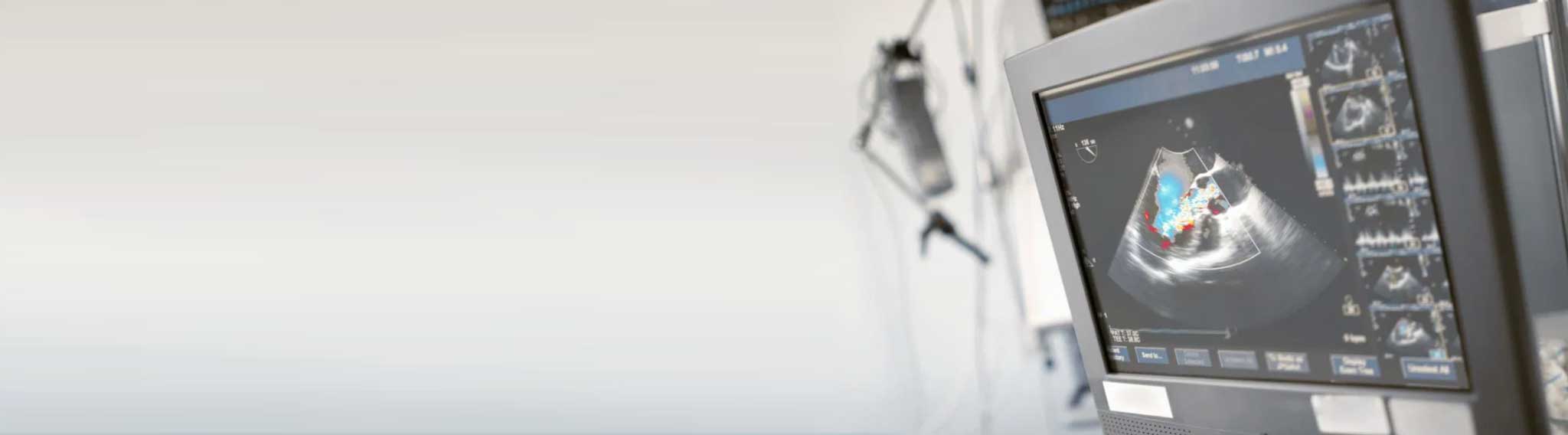
ECHO Cardiogram (2D/3D ECHO)
Ultrasound Scan of the Heart (Non-Invasive, No Pain)
Echocardiography, also known as cardiac ultrasound, uses ultrasound to create real-time images of the heart. It is painless, non-invasive, and highly effective in assessing heart structure and function.
What Is Echocardiography?
Echocardiography provides detailed insights into the heart’s size, shape, pumping strength, and valve function. It also helps detect tissue damage, calculate cardiac output, ejection fraction, and evaluate how well the heart relaxes (diastolic function).
Why Is It Done?
Echocardiography plays a crucial role in diagnosing and managing heart conditions:
- Early diagnosis of myocardial infarction (heart attack) by showing wall motion abnormalities
- Monitoring and treatment of heart failure by assessing ejection fraction
- Detection of cardiomyopathies like hypertrophic or dilated cardiomyopathy
- Using stress echocardiography to investigate chest pain and related symptoms
Doppler & Advanced Imaging
Doppler echocardiography evaluates blood flow through the heart using pulsed or continuous wave techniques. It helps assess valve function (regurgitation or stenosis), detects abnormal flow between heart chambers, and measures tissue velocity using tissue Doppler.
Types of Echocardiography
- Trans-thoracic Echocardiogram (TTE): The most common type. It uses a probe placed on the chest wall to view the heart. It is non-invasive and provides quick, accurate assessment.
- Trans-esophageal Echocardiogram (TEE): A specialized probe is inserted via the mouth into the esophagus to get a closer and clearer image of the heart’s structures.
TTE Imaging Windows
Multiple imaging windows are used in TTE for detailed assessment:
- Parasternal long and short axis views
- Apical two, three, and four chamber views
- Subcostal window (beneath the ribcage)
These may be displayed in M-mode (one-dimensional), 2D, or 3D formats and are often combined with Doppler techniques for full evaluation.
Advanced Features at Virgo Heart Center
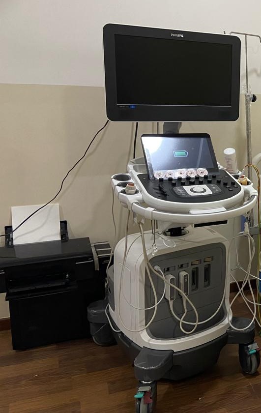
At Virgo Heart Center, we use the AI-enabled Philips Affiniti 70 Echo Machine with 2D and 3D capabilities. Our team is highly trained in advanced echo techniques including:
- Speckle Tracking and Global Longitudinal Strain (GLS)
- Stress Echocardiography for detecting ischemic heart conditions
Echocardiography is safe, non-invasive, and has no known risks, making it an essential tool in modern cardiology.